Third molar extraction, popularly known as the removal of wisdom teeth, is a common procedure performed by dentists specializing in oral and maxillofacial surgery.
These teeth, which usually appear between the ages of 17 and 25, can cause a number of problems when there isn’t enough space in the mouth to accommodate them properly or when they are associated with the presence of a cyst.
There are several reasons why wisdom teeth removal may be recommended:
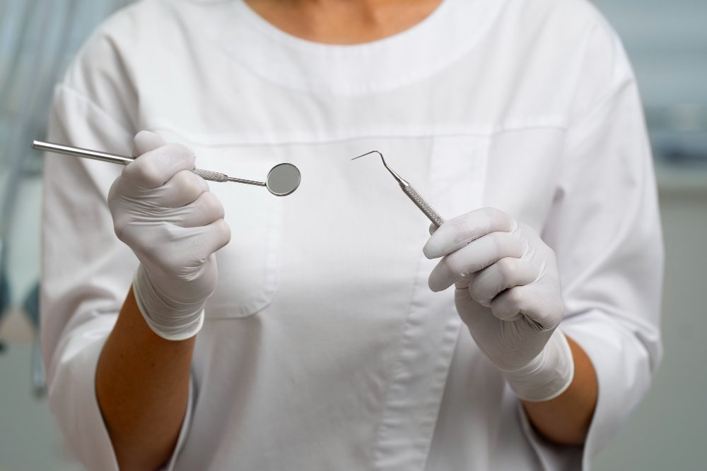
The removal of third molars is a relatively straightforward procedure that can usually be performed under local anesthesia and conscious sedation.
On some occasions it may need to be performed under general anesthesia, depending on the complexity of the extraction, but this is a very infrequent situation.
After wisdom teeth extraction, it is important to follow the guidelines to ensure a smooth recovery:
If you notice any of the following signs after the extraction, contact your dentist immediately:
Third molar extraction is a common and safe practice when performed by qualified professionals.
By following the guidelines and post-operative care, most patients recover quickly and without significant complications.
If you have any questions or concerns, don’t hesitate to talk to our team.
Orthodontic mini-plates are bone anchoring devices that allow orthodontists to make orthodontic mechanics more efficient.
As a result, it is now possible to correct malocclusions without having to extract premolars in most cases, and in many cases avoiding the need for orthognathic surgery.
The miniplates are installed through a small incision in the gum under local anesthesia, through which the miniplate is fixed to the bone.
After orthodontic treatment, the device is removed through the same place as the previous incision.


Guided Bone Regeneration is a technique used in implantology and oral and maxillofacial surgery to promote bone growth in specific areas, establishing a solid base for the placement of dental implants.
This procedure involves the use of materials such as membranes and biomaterials to direct bone growth in a controlled manner.
These materials act as physical barriers, preventing the growth of soft tissue and allowing bone to regenerate in the desired area.
Guided Bone Regeneration is indicated to treat bone defects, increase bone thickness or height and prepare the site for the insertion of dental implants.
The lip brakes have the function of limiting lip movements, stabilizing the midline and preventing excessive exposure of the gums.
The lingual brake, on the other hand, limits the movement of the tongue, facilitating swallowing, speech and phonation.
In some cases, alterations can occur in the function of the brakes, resulting in a pathological lip brake.
This can lead to the presence of interincisal diastema (space between the teeth), aesthetic problems, biofilm build-up, abnormal traction of the upper lip, difficulty in brushing and problems with the phonetics of some letters.
Procedures such as frenotomy and frenectomy are performed to correct these brake disorders, whether labial or lingual.
Frenectomy: A frenectomy is a surgical procedure designed to remove the labial and lingual bridles.
This procedure allows both orthodontic movement to close diastemas and adequate tongue movement, which is essential for functional activities.
Canine fenestration is a dental procedure that aims to help align canine teeth that have not erupted correctly.
This procedure is commonly carried out on orthodontic patients where the canines are impacted, i.e. stuck inside the jawbone or gums and cannot emerge naturally.
Fenestration is a surgical procedure in which gum tissue and sometimes bone is removed to expose the impacted canine tooth.
This allows the orthodontist to apply force to move the tooth into the correct position in the dental arch using braces.
After canine fenestration, it’s essential to follow all of Dr. G.’s guidelines to ensure a smooth recovery.
to ensure a smooth recovery:
Dental implants are titanium components that act as the root of a tooth and are placed through surgery in the jawbone, below the gum.
The three main components of a dental implant are:
After a short period of time, the implant integrates with the bone, providing stable support for the artificial teeth that are placed on these implants.
Dental implants are also beneficial for general oral health because, unlike bridges, they don’t need to be attached to another tooth and they also stop the process of resorption of the jawbone that begins when a tooth is lost.
Although there are many types of implants: cylindrical, conical… the main distinction is between three types, based on where they are placed:
Different types of dental implants can be combined to fix a prosthesis and obtain better results.
Orthognathic surgery is an advanced procedure performed to correct irregularities in the jaw and maxillary bones.
This type of surgery is indicated for patients who have problems such as facial asymmetry, open bite, retrognathia or mandibular prognathism, among others.
As well as improving facial aesthetics, orthognathic surgery can relieve pain and functional problems, such as difficulties chewing, speaking and breathing.
Patients with this type of problem usually have a dento-skeletal alteration, which means that the position of their teeth and jaws needs to be corrected.
For this reason, most patients need prior orthodontic treatment in order to undergo orthognathic surgery with the best possible results.
Planning orthognathic surgery involves a 3D approach to ensure precision and better results.
With this technology, it is possible to simulate the surgery before the procedure, offering a clear visualization of the final result.
The benefits include not only a more harmonious appearance, but also a significant improvement in masticatory and respiratory function.
There are three types of orthognathic surgery:
Orthognathic jaw surgery aims to correctly position the jaw in order to achieve facial harmony and, above all, to restore the functionality of this bone, which is essential for chewing, breathing and speaking.
This procedure corrects facial deformities such as mandibular prognathism (Class 3), open bite and gummy smile.
It consists of a Le Fort I osteotomy, which allows the jawbone to be advanced, retracted, lengthened, shortened or rotated.
The bone is fixed with biocompatible titanium plates.
Due to the risks of mandibular retraction surgery (narrowing of the airway), the most common orthognathic surgery on the mandible is mandibular advancement.
This procedure is necessary in cases where the jaw is small and retracted in relation to the maxilla, a condition known as retrognathia or Class II.
In addition to its undeniable aesthetic benefits, this surgery has a huge functional impact as it increases the width of the airway and is the gold standard treatment for problems such as obstructive sleep apnea.
The surgery is performed inside the mouth, so there is no visible incision outside the mouth.
A cut is made through this incision so that the mandible can be fixed in the correct position using fully biocompatible titanium plates.
In cases where the patient needs correction of the positioning of both the maxilla and mandible, a bimaxillary surgery is planned in which both the upper and lower parts are corrected.
The surgery takes place in a hospital under general anesthesia.
Depending on the extent of the surgery, the patient usually stays in hospital for one day and is then released to continue recovering at home.
During the post-operative period, the patient should follow the doctor’s instructions, eat a liquid diet for 30 to 45 days and start receiving care from a specialized physiotherapist.
Copyright © Irene Méndez-Manjón | Developed by Libelle Comunicação
Fale Conosco